Our Health Library information does not replace the advice of a doctor. Please be advised that this information is made available to assist our patients to learn more about their health. Our providers may not see and/or treat all topics found herein.
Electrocardiogram (EKG) Components and Intervals

An electrocardiogram (EKG, ECG) is a test that measures the electrical signals that control heart rhythm. The test measures how electrical impulses move through the heart muscle as it contracts and relaxes.
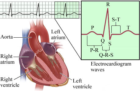
The electrocardiogram translates the heart's electrical activity into line tracings on paper. The spikes and dips in the line tracings are called waves.
- The P wave is a record of the electrical activity through the upper heart chambers (atria).
- The QRS complex is a record of the movement of electrical impulses through the lower heart chambers (ventricles).
- The ST segment shows when the ventricle is contracting but no electricity is flowing through it. The ST segment usually appears as a straight, level line between the QRS complex and the T wave.
- The T wave shows when the lower heart chambers are resetting electrically and preparing for their next muscle contraction.
Current as of: May 1, 2025
Author: Ignite Healthwise, LLC Staff
Clinical Review Board
All Ignite Healthwise, LLC education is reviewed by a team that includes physicians, nurses, advanced practitioners, registered dieticians, and other healthcare professionals.
This information does not replace the advice of a doctor. Ignite Healthwise, LLC disclaims any warranty or liability for your use of this information. Your use of this information means that you agree to the Terms of Use and Privacy Policy. Learn how we develop our content.
To learn more about Ignite Healthwise, LLC, visit webmdignite.com.
© 2024-2026 Ignite Healthwise, LLC.


